
therefore, neurons with lower axial resistance (larger diameter) will have longer length constants Neurotransmitters are released by cells near the dendrites, often as the end result of their own action potential These incoming ions bring the membrane.axial resistance is the internal resistance to flow: the larger the diameter of the structure, the more opportunity there is for the ions to flow.

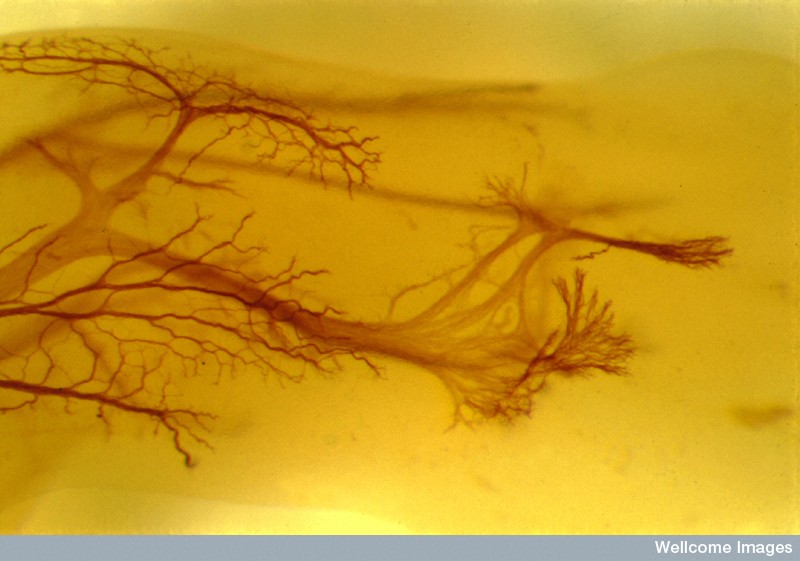
Dendrites function to receive information, and do so through numerous. therefore, neurons with higher membrane resistance (fewer open channels) will have longer length constants Dendrites: Dendrites are short, branched processes that extend from the cell body.individual cells, and even different patches of membrane within a given cell, will have different length constants because the length constant is dependent upon the membrane resistance and the axial resistanceĬan be thought of as "leakiness": as the ions that cause the V m change move away from their source, they can move back through the membrane via other open channels.this decay can be described by the length constant (λ), which corresponds to the distance where a graded potential has decreased to 37% of its original amplitude.in the absence of any active, regenerative processes (see action potentials), a graded potential will decay as it travels away from its site of origin The prevalence of such failures of action potential backpropagation depend on the biophysical properties and morphology of the dendritic arbor.The membrane potential will begin at a negative resting membrane potential, will rapidly become positive, and then rapidly return to rest during an action potential. The change in the membrane voltage from -70 mV at rest to +30 mV at the end of depolarization is a 100-mV change. The action potential is a brief but significant change in electrical potential across the membrane. Transmission of a signal within a neuron (from dendrite to axon terminal) is carried by a brief reversal of the resting membrane potential called an action. It is the electrical signal that nervous tissue generates for communication. membrane voltage changes occur both in time and in space (more about this under synaptic integration) What has been described here is the action potential, which is presented as a graph of voltage over time in Figure 12.5.7.


 0 kommentar(er)
